
waste water treatment plant angel chemical cationic at malaysia

Our company was founded in 2002, Mainly engaged in polymer series products, it is a modern high-tech enterprise integrating R&D, production, sales and application services. At present, there are more than 100 kinds of research and development products, which are mainly sold to many countries and regions…
hottest sellingwater treatment chemicals – aquasol at thailand
Silver staining is used to detect proteins after electrophoretic separation on polyacrylamide gels. It combines excellent sensitivity (in the low nanogram range) whilst using very simple and cheap equipment and chemicals. It is compatible with downstream processing such as mass spectrometry analysis after protein digestion.
Silver staining of proteins in polyacrylamide gels
The silver-staining procedure for detecting proteins in polyacrylamide gels has been modified and further simplified so that it is stable, controllable, and even more rapid than previous silver-staining methods. The method retains its sensitivity to proteins at the nanogram level and may be used either before or after Coomassie blue staining.
Get Price
Silver staining of proteins in polyacrylamide gels.
Silver staining of proteins in polyacrylamide gels. Chevallet M(1), Luche S, Rabilloud T. Author information: (1)CEA-Grenoble, DRDC/ICH, 38054 Grenoble, Cedex 9, France. Silver staining is used to detect proteins after electrophoretic separation onpolyacrylamide gels. It combines excellent sensitivity (in the low nanogramrange) with the use of very simple and cheap equipment and chemicals.
Get Price
Silver staining of proteins in polyacrylamide gels
Silver staining is used to detect proteins after electrophoretic separation on polyacrylamide gels. It combines excellent sensitivity (in the low nanogram range) with the use of very simple and
Get Price
Modified method of silver staining of proteins
The very sensitive and reliable silver staining method to visualize proteins in polyacrylamide gels described by Wray et al. (Anal.Biochem. (1981) 118, 197–203) fails when the protein sample contains nucleic acids and/or metals.By washing the polyacrylamide gels in acetic acid and repeatedly in methanol immediately following electrophoresis and then using the procedure of Wray et al., many
Get Price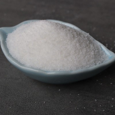
Silver staining of proteins in polyacrylamide gels.
polyacrylamide gels. It combines excellent sensitivity (in the low nanogram range) whilst using very simple and cheap equipment and chemicals. It is compatible with downstream processing such as mass spectrometry analysis after protein digestion. The sequential phases of silver staining are protein fixation, then sensitization, then
Get Price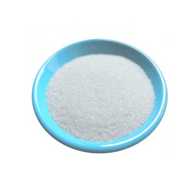
Silver staining of proteins in polyacrylamide gels: a
The introduction of silver staining of proteins in polyacrylamide gels in 1979 (Switzer et al. 1979) has been a breakthrough in the field of protein detection, raising the sensitivity from the microgram range obtained with dyes such as Coomassie blue to the nanogram range. This
Get Price
Silver staining DNA in polyacrylamide gels | Nature Protocols
This protocol describes a simple silver staining method used to visualize DNA fragments and other organic molecules with unsurpassed detail following traditional polyacrylamide gel electrophoresis
Get Price
Silver staining of proteins in polyacrylamide gels.
polyacrylamide gels. It combines excellent sensitivity (in the low nanogram range) whilst using very simple and cheap equipment and chemicals. It is compatible with downstream processing such as mass spectrometry analysis after protein digestion. The sequential phases of silver staining are protein fixation, then sensitization, then
Get Price
Title: Silver-staining of proteins in polyacrylamide gels
Abstract: On the basis of the physico-chemical principles underlying silver-staining of proteins, which are recalled in this paper, several methods of silver-staining of proteins after SDS electrophoresis in polyacrylamide gels or isoelectric focusing were tested. The most valuable protocols are presented in this report, including standard methods for unsupported gels and new methods devised
Get Price
Silver Stains for Protein Gels - Fisher Scientific
Silver Stains for Protein Gels Thermo Scientific™ Pierce™ Silver Stain Kit Stain proteins in 1D and 2D gels with this rapid, ultra-sensitive and versatile silver stain system that yields consistent and reliable results.
Get Price
Simplified method for silver staining of proteins
The study of effects of several parameters on silver staining of proteins has led to the development of a staining method which is simple and reliable, requires only few stable solutions, and can be applied to all gel types such as sodium dodecyl sulfate (SDS) containing polyacrylamide or isoelectric focusing gels.
Get Price
Fluorescent Silver Staining of Proteins in Polyacrylamide Gels
Silver staining is a colorimetric technique widely used to visualize protein bands in polyacrylamide gels following sodium dodecyl sulfate-polyacrylamide gel electrophoresis (SDS-PAGE).
Get Price
High-Sensitivity Silver Staining of Proteins Follo
High-Sensitivity Silver Staining of Proteins Following Polyacrylamide Gel Electrophoresis: Polyacrylamide gel electrophoresis is a simple, inexpensive, yet highly versatile and powerful method for the analysis of complex mixtures of proteins. In part, the success of this method has resulted from the ease with which the fractionated proteins can
Get Price
PlusOne Silver Staining Kit, protein | Cytiva, formerly GE
Silver Staining Kit, Protein provides convenient, reproducible and sensitive staining of proteins in polyacrylamide gels .including PhastGels in less than two hours The visualization technique detects most proteins in the nanogram range, which is 100-fold more sensitive than Coomassie Blue staining.
Get Price
Silver Staining Kit, Protein GE Healthcare, 17-1150-01
PlusOne Silver Staining Kit, Protein and PlusOne DNA Silver Staining Kit are two silver staining kits that bring speed, convenience, and very high sensitivity to the staining of proteins and nucleic acids in polyacrylamide electrophoresis. gels. Silver Staining Kit, Protein includes detailed protocols and components in quantities sufficient to
Get Price
Title: Silver-staining of proteins in polyacrylamide gels
Abstract: On the basis of the physico-chemical principles underlying silver-staining of proteins, which are recalled in this paper, several methods of silver-staining of proteins after SDS electrophoresis in polyacrylamide gels or isoelectric focusing were tested. The most valuable protocols are presented in this report, including standard methods for unsupported gels and new methods devised
Get Price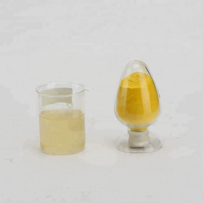
Silver stain for proteins in polyacrylamide gels: A
The rapid, ultrasensitive silver stains that have been developed recently for detecting proteins in polyacrylamide gels show variation in staining from gel to gel and do not stain certain proteins at all.
Get Price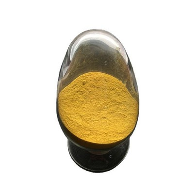
Fluorescent Silver Staining of Proteins in Polyacrylamide Gels
Silver staining is a colorimetric technique widely used to visualize protein bands in polyacrylamide gels following sodium dodecyl sulfate-polyacrylamide gel electrophoresis (SDS-PAGE).
Get Price
The mechanism of silver staining of proteins separated
abstract = "Gel based silver staining of proteins is thought to occur by selective reduction of silver ions to insoluble metallic silver at specific initiation sites in the vicinity of the protein molecules.
Get PriceImproved silver staining of plant proteins, RNA and DNA
The performance of this method is documented by staining one‐and two‐dimensional patterns of plant leaf proteins. Moreover, we achieved, for the first time, the detection of the non‐structural, tobacco mosaic virus‐specific 126 kDa protein directly in the one‐dimensional protein pattern of infected protoplasts by a staining procedure.
Get Price
Silver Stain Plus Kit | Life Science Research | Bio-Rad
Silver staining is a highly sensitive method for detecting proteins and nucleic acids in polyacrylamide slab gels. Bio-Rad silver stain kits are 10–50-fold more sensitive than Coomassie Brilliant Blue R-250 for proteins (detection is ~0.1 ng/mm 2) and 2–5-fold more sensitive than ethidium bromide for single and double-stranded DNA and RNA.. Silver Stain Plus™ stain is the most sensitive
Get Price
A Highly Sensitive Silver Stain for Detecting Proteins
Ultrasensitive silver-stain method for the detection of proteins in polyacrylamide gels and immunoprecipitates on agarose gels. Porro M, Viti S, Antoni G, Saletti M. Porro M, et al. Anal Biochem. 1982 Dec;127(2):316-21. doi: 10.1016/0003-2697(82)90179-8.
Get Price
Silver Stains for Protein Gels
Antibodies & Protein Biology Antibody Production & Purification; Electrophoresis, Western Blotting and ELISA
Get Price
Silver Staining – Destaining Kit | Krackeler Scientific, Inc.
Amresco's Silver Staining – Destaining Kit is a combination of two products for convenience; Silver Bullit™ Silver Stain Kit (for 50 mini-gels) and Silver Subtract™ Silver Destaining Reagent (for 12 mini-gels). Proteins can be detected in polyacrylamide gels with high sensitivity and nearly undetectable background using Amresco’s Silver
Get Price
Improved silver staining of plant proteins, RNA and DNA
The performance of this method is documented by staining one‐and two‐dimensional patterns of plant leaf proteins. Moreover, we achieved, for the first time, the detection of the non‐structural, tobacco mosaic virus‐specific 126 kDa protein directly in the one‐dimensional protein pattern of infected protoplasts by a staining procedure.
Get Price
Rapid Staining of Proteins in Polyacrylamide Gels
Abstract: Sodium dodecyl sulfate-polyacrylamide gel electrophoresis (SDS-PAGE) is one of the most powerful methods for protein analysis. Unfortunately, the typical procedures for the detection of protein bands after SDS-PAGE, using the visible dye Coomassie blue and silver staining, have several time-consuming steps and require the fixation of proteins in the gel.
Get Price
Mass Spectrometric Sequencing of Proteins from Silver
(NanoES),21,22 which we have used for the sequencing of proteins from polyacrylamide gels.23 In the course of that work, it became apparent that the 50-100 ng detection limit of the Coomassie staining method was not low enough for the levels that could be reached. Silver staining is a popular and more sensitive staining method,
Get PriceGE Healthcare PlusOne™ Silver Staining Kit for Protein
Description: PlusOne Silver Staining Kit, Protein and PlusOne DNA Silver Staining Kit are two silver staining kits that bring speed, convenience, and very high sensitivity to the staining of proteins and nucleic acids in polyacrylamide electrophoresis. gels.
Get PricePurify proteins from polyacrylamide gels
gel to determine which section of the unstained gel should be excised and 2) stain the entire gel with a negative stain or other type of stain that can be reversed after excising the band. The second step in purifying electrophoresed protein from polyacrylamide gels is to extract (elute) the protein from the gel matrix.
Get PriceRelated Products


